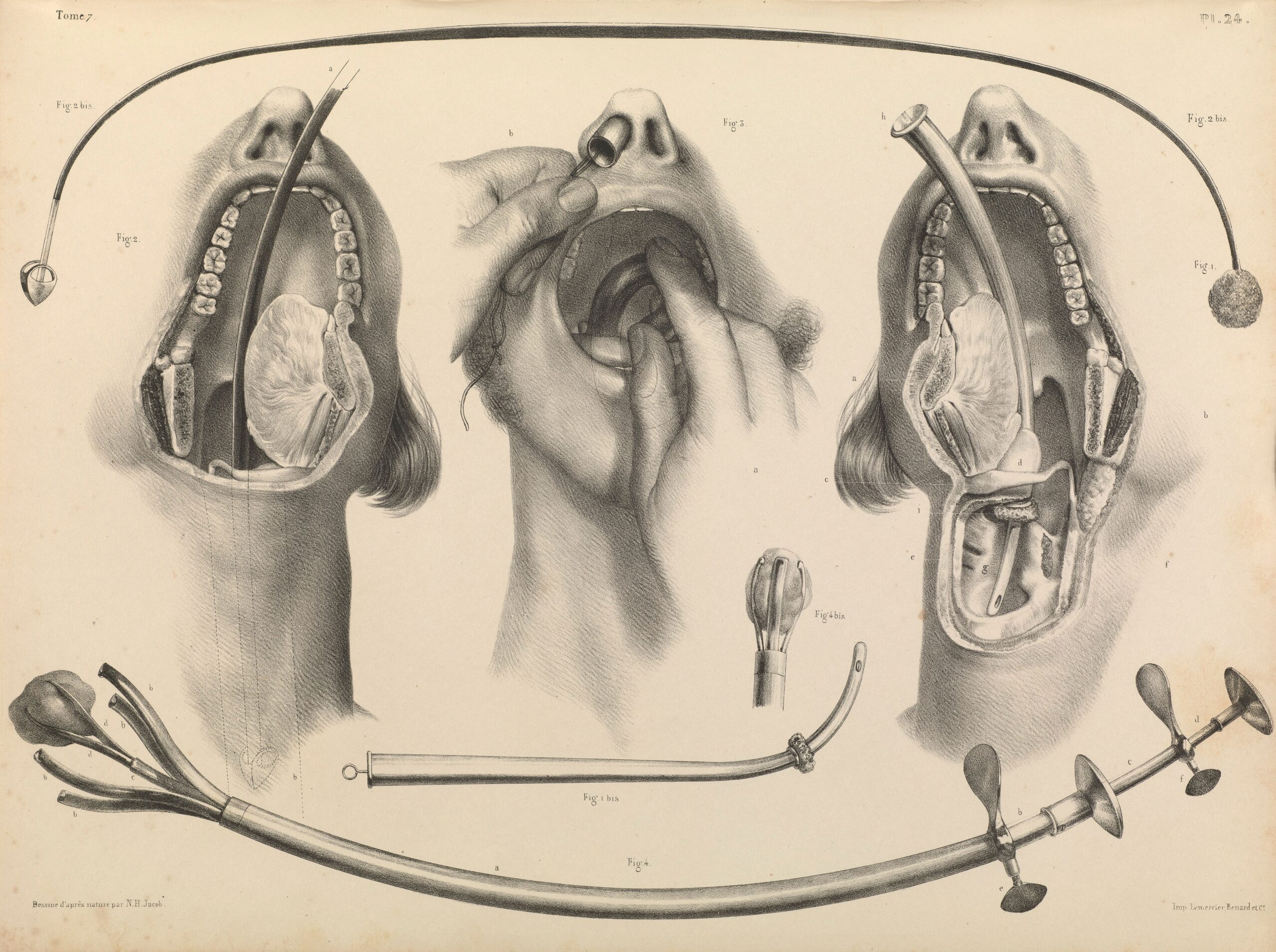
The 19th century saw advancements in dental illustration via the advent of lithography and photography. Lithography involves drawing on a flat surface with an oil-like substance and then making prints from it by pressing paper onto the oil. This allows for the mass production of detailed illustrations. This technology allowed illustrations to be printed in textbooks and journals in a more cost-efficient manner than the use of woodcuts with the printing press, significantly increasing who could publish and access this anatomical knowledge.
At the same time, the development of photography introduced a new level of realism to all forms of visual documentation. Photographs could be used as references for, or alongside, illustrations to document dental conditions and procedures with a new level of accuracy. As photograph technology, both in terms of the cameras and print creation, became more advanced and user-friendly, photos became a valuable and more utilized asset for creating educational resources.
As dental practices and technologies evolved throughout the 20th century, so did the tools and techniques of dental illustrators. Starting in the 1990’s, the use of digital illustration, 3D models, and 3D animations increased the scope of what dental illustration can accomplish. Digital illustration techniques make work infinitely reproducible and significantly increase the speed with which illustration can be created. 3D modeling software allows for illustrations that show a subject matter at any angle, perspective, or in any context. While 3D animation has allowed these illustrations to move, removing any ambiguity caused by misunderstandings of diagrams or infographics. 3D animations can walk viewers through exactly what the illustrator wants to be shown, as well as having audio narration to further help the viewer comprehend what is being shown. Animations, while being slightly less accessible or widely distributed, can deliver information to dental professionals and patients in ways that are much more easily understood than drawn-out text based descriptions, or 2D illustrations.
Today, dental illustration continues to play an important role in educating dental professionals, informing patients, and advancing research: A reflection of the ongoing evolution of both the art and the science in dental care.


