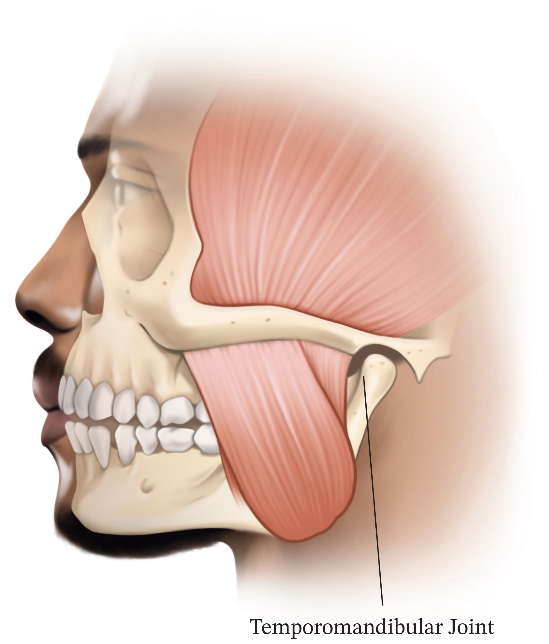
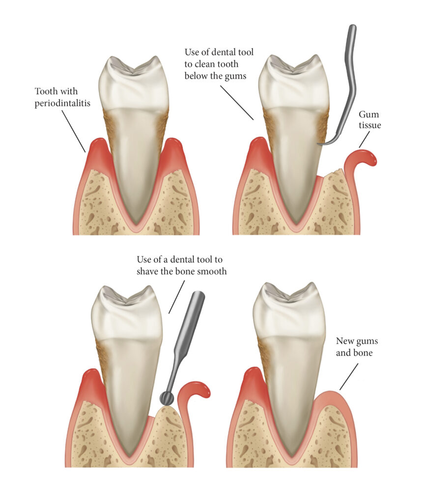
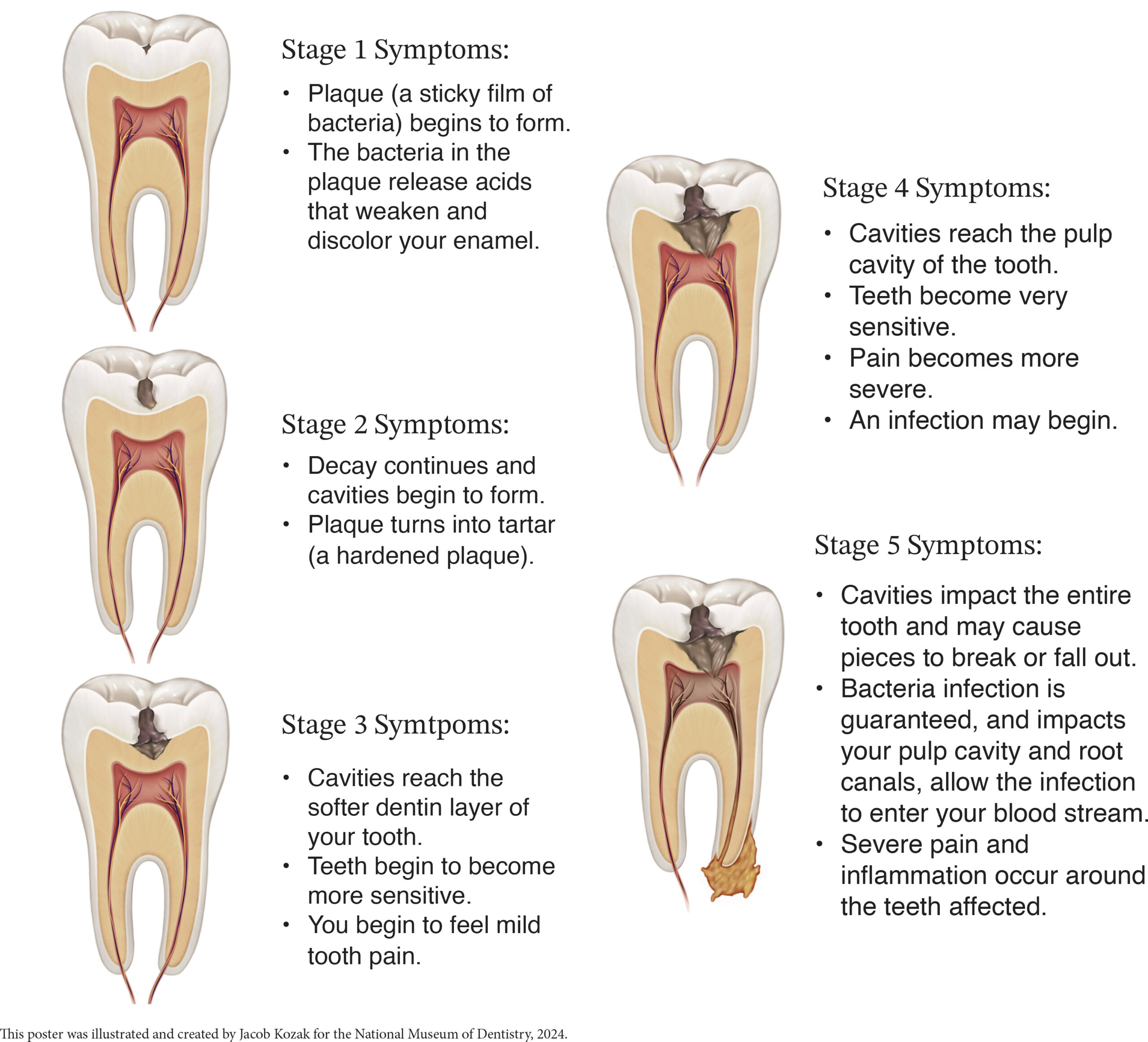
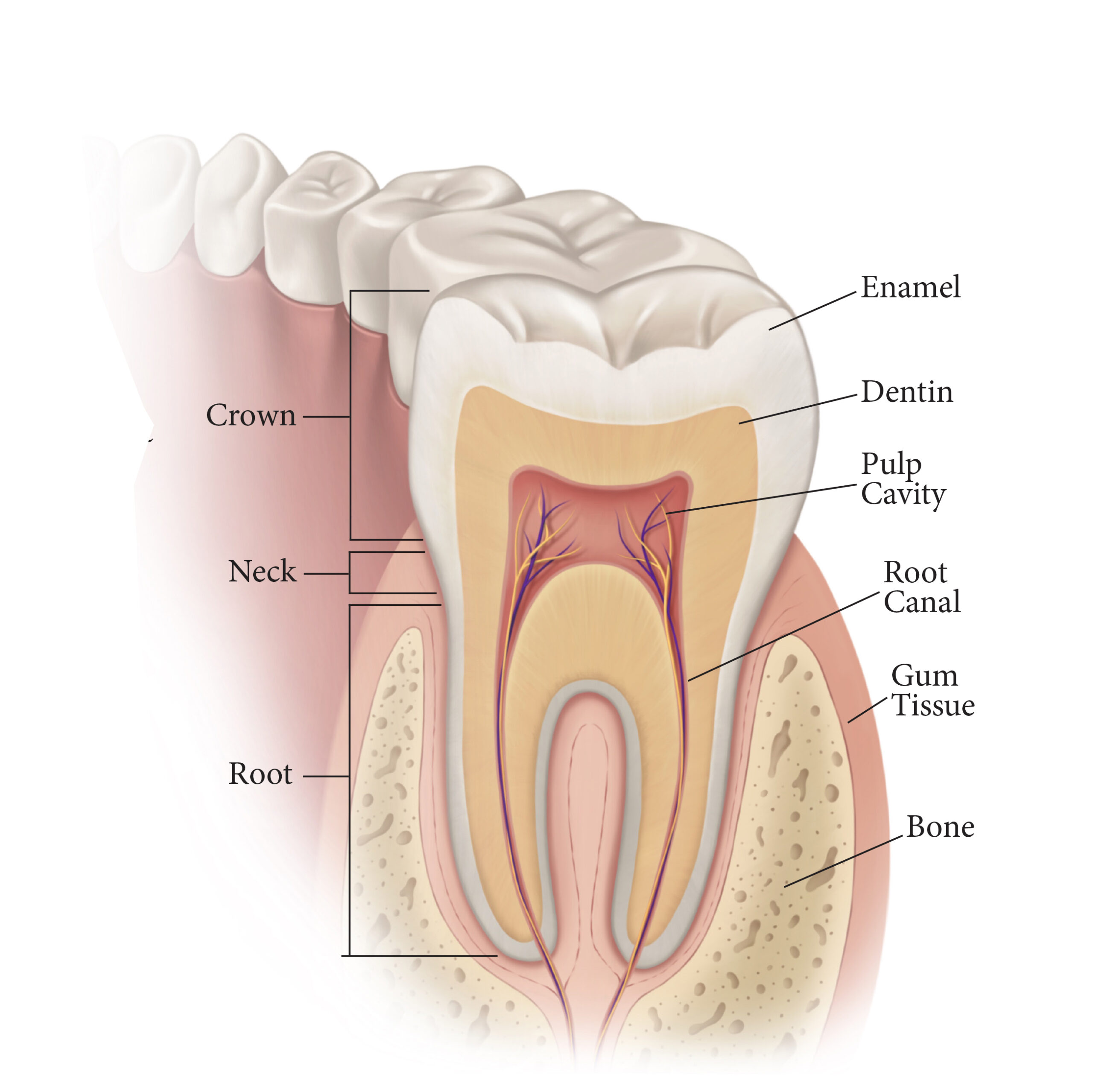
Dental illustration creates high-quality, detailed representations of the human body, including muscles, bones, and important anatomical structures that can be difficult to explain using only words.
Illustrations can be used to teach students about structure and function, and can commonly be found in textbooks and lectures as visual aids to complement the information being presented. Illustrations can also show the effects of diseases, conditions, and injuries on the body.
These can include visualizations of tumors, infections, and other abnormalities not readily observable on the surface of the human body. More advanced illustrative tools often include interactive 3D models, virtual dissections, and animated diagrams. These digital resources allow students to explore the anatomy dynamically as opposed to in two-dimensions. Detailed drawings or digital animations of surgical procedures provide a step-by-step visual guide, helping students understand complex techniques and surgical anatomy. Some educational programs use medical illustrations with simulation tools to create realistic training environments. These simulations may include virtual reality (VR) or augmented reality (AR) components that integrate illustrative visuals with advanced computer technology.
The use of illustration in dental education is well documented. According to the University of New South Wales, Australia, in their Open Access Journal of Dental and Oral Surgery (OAJDOS), Visual learning is important in education and learning. Based on the review findings of the article “Online Illustrations for Students’ SelfLearning: A Review using Dentistry as an Example” students found that online illustrative material was important, helpful and enhanced confidence in their learning. The illustrations were most useful as supplemental learning tools, and were especially well received when used to aid in teaching simple procedures. This reaffirms the importance of including pictures and illustrations during learning courses.
Dental and Medical illustrations are not only used to accurately convey subjects, but also to simplify complex information. When surveyed, students expressed that simplified graphs and representational illustrations, in conjunction with text elements, were their preferred way of learning about new concepts. Of course, the effectiveness of dental illustrations is dependent on many factors, and there is no single, perfect way of delivering information. Variations in style, perspective, and level of detail can lead to different interpretations or misunderstandings of an image. That is why it is important that these illustrations be accompanied by other methods of teaching, so as to provide a more complete understanding of complex medical concepts.


